


- HOME
- For Patients
- Glioma
- Surgical treatment under the MRI
Glioma
- About glioma
- Symptom of glioma
- Diagnosis of glioma
- Treatment of glioma
- Surgical treatment under the MRI
Surgical treatment under the MRI

Tokai University has further secured its worldwide technological leadership in the medical field with the installation of an interventional suite in the University Hospital in Kanagawa, Japan, in February 2006.

Magnetic resonance/X-ray/operation suite (MRXO) is a seamless integration of Magnetic resonance imaging (MRI), computed tomography (CT), and X-ray imaging with a high-end operating table in a futuristic and highly efficient approach to image-guided surgery.


The installation was the result of a special project requested by Professor Mitsunori Matsumae, the Neurosurgeon-in-Chief and Chair of the Department of Neurosurgery at the Tokai University School of Medicine. The design and installation of the MRXO suite was achieved with the combined efforts of the Philips Medical Systems Magnetic Resonance, Cardiovascular X-ray, and Computed Tomography Business Groups, in close collaboration with Professor Matsumae. Mizuho Ika Kogyo in Japan developed the surgical table, while Toda Co. Ltd. undertook the construction work at Tokai University Hospital.
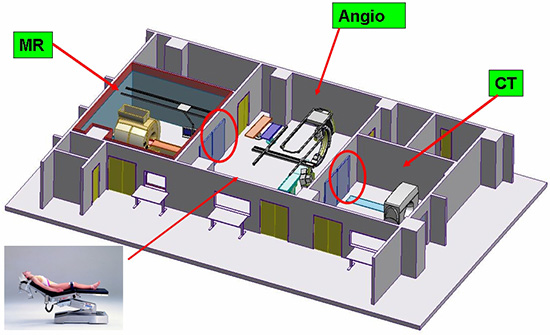
CAPTION: Schematic diagram of Tokai University's MRXO setup. Left: MR scanner, center: cath lab combined with operation room, right: CT scanner. Electric sliding doors enable either combined or single use of the separate areas.
The suite is equipped with ideally-positioned radiological equipment (CT, MRI, and angiography) combined with a fully-functional operating room. The unique layout of this "operating room of the future" allows a patient, who is positioned in the centre of the suite, to be transported to MRI, CT, or X-ray diagnostic systems within minutes; the mobile table top carrying the patient is easily transported between the different imaging modalities and the operating table, while the most appropriate modality can be selected at any particular moment during any surgical procedure. The MRI and CT rooms can also be closed with sliding doors so that all imaging systems can be used independently, enabling unsurpassed flexibility and economic efficiency.


The treatment room is the core of the suite; it contains angiographic equipment within a fully functional operating room. The tabletop enables both examination and treatment, particularly surgical procedures. The most significant feature of this system is the ease of patient transfer between examinations during treatment, e.g., from angiographic examination to MRI or CT without changing beds. The patient can be transferred easily from the operating table to the MRI, and back to imaging once again. Patient transfer during treatment is performed with the utmost care under the direction of a specially trained safety supervisor.

The core treatment room is equipped with an operation room (OR)-grade clean air-conditioning system, operating lights, medical gas outlets, and an in-hospital information system; this enables various surgical procedures to be undertaken, including craniotomy.
The adjacent CT and MRI rooms are also equipped with medical gas outlets, an OR-grade clean air-conditioning system, and operating lights, etc., for the safe transfer of an anesthetized patient to CT or MRI for diagnosis.

Each of the imaging systems in isolation is capable of performing diagnostic examinations of the whole body; however, once treatment (for example, surgery) commences in the OR located in the center of the suite, imaging is easily performed as required to provide intraoperative information using high-performance diagnostic equipment.

Surgery for intracranial disease aims to accomplish safe treatment with high precision in a short period of time. Here, the key that governs the progress of the operation is the intraoperative identification of the extent of preoperative planning achieved at a given time. We consider that this suite enables precise patient treatment within a shorter time period than previously possible because the progress of the operation can now be identified using intraoperative imaging as required. In practical terms, the system facilitates intraoperative patient transfer from the treatment room to the MRI examination room for verification of the patient's condition and the degree of procedural progress before returning to the treatment room and return transfer to the examination room for final verification before the operation is completed; surgery will proceed more smoothly and safely than previously possible.

The advantages of the MRXO system are utilized in the following procedures:
- Diagnosis and treatment of stroke
- Neurosurgical procedures
- Treatment of intra-abdominal tumors
- Treatment of any vascular occulted disease
- Routine diagnosis of a variety of diseases
Operating rooms equipped with CT scanner, MRI, or angiographic equipment have been developed previously; however, this newly-built MRXO suite at Tokai University Hospital is a more evolved, integrated system of radiological diagnosis/treatment and surgery that represents a "neo-futuristic operating room". Tokai University Hospital will endeavor to enhance the efficacy of treatment by actively utilizing this MRXO suite.

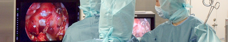
Professor Matsumae performed the first surgical procedure in the suite in February 2006 on a patient with a large left-sided brain tumor. The MR scanner was used to check progress at several stages during tumor resection by quickly transferring the patient from the operating table into the MR scanner. This modality was also used to check for complications, to ensure that all tumor tissue had been removed, and to detect any damage to healthy brain tissue that might lead to major complications for the patient. The world's first MRXO intervention case was accomplished successfully.

One or two cases of neurosurgery and two cases of radiological intervention are currently performed per week. In addition, an average of 40 diagnostic CT cases, 16 diagnostic MRI cases, and one angiography case are performed each day using these facilities, both day and night.
The MRXO suite was planned by the Tokai University School of Medicine and completed with the cooperation of Philips Medical Netherlands Headquarters, Electronics Japan Medical Systems, Toda Co. Ltd., and Mizuho Ika Kogyo Co. Ltd.

Team of the MRXO in Tokai University Hospital

Team of the Philips Medical Systems

Team of the Mizuho Ika Kogyo
This highly successful MRXO project is another milestone for innovation in medical technology and reflects the flexible attitude of Philips with respect to the needs of our patients.
