


- HOME
- For Patients
- Brain tumor
- Standard treatment
Brain tumor
Standard diagnostic method
Surgery is the basic treatment for brain tumor
In some conditions, tumors may remain completely asymptomatic, or remain stable without progression even though some symptoms are present. In such cases, the course of the condition may be monitored by outpatient MRI without performing surgery. Conditions that are sufficiently severe to produce symptoms due to intracranial hypertension, however, or of types that can be anticipated to progress on the basis of test results despite the presence of relatively minor symptoms, are treated using a range of methods. The method of treatment varies depending on the type of disease in question, but surgery is the most powerful method available.
Surgery has two purposes
1.To enable accurate assessment of the disease
It may not always be possible to tell what type of tumor is present from MRI and other tests. In this case, a test called "pathological examination" is carried out to assess whether it is safe just to keep the tumor under observation or whether treatment such as radiotherapy or drugs should be used, and if so, which drug to choose. Pathological examination involves using a microscope to examine a tumor that has been surgically removed in order to determine its type.
2.To remove the disease
The basic purpose of surgery is to cure the disease by removing the tumor. When establishing a treatment plan, however, a number of important points are always considered prior to surgery. For patients with conditions requiring surgery:
- The disease cannot be excised, including healthy (normal) areas;
- In many cases, the borders of brain tumors are unclear, and it is difficult to tell how large an area needs to be excised with the naked eye;
- Important blood vessels, nerves, and the surrounding brain should not be damaged in order to observe or excise the tumor.
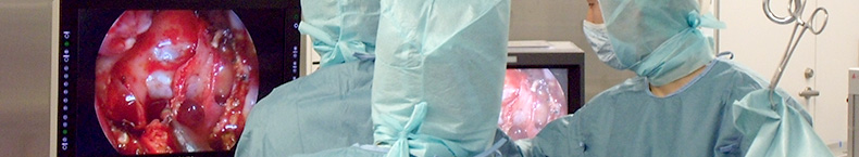
That is, a variety of surgical techniques have been developed in order to remove as much of the tumor as possible without causing after-effects following surgery. At Tokai University Hospital, we use a technique called MRXO to carry out surgery while observing an image of the tumor onscreen.
What is MRXO intraoperative diagnostic imaging?
During an operation, surgeons are always careful about what is at the end of their scalpel. While remaining focused on a small area of disease, they also have to think about the position and order of healthy structures. Areas covered by the diseased part and deeper areas (depth and trajectory of cutting), however, are extremely difficult to assess. This can be a barrier to the smooth progress of surgery, as the surgeon carrying out the operation must remember the images (CT or MRI scans) taken prior to surgery and match their memory with the actual scene in accordance with the progress of the operation.
Advances in diagnostic imaging and computer technology in recent years have enabled the development of navigation equipment for use during surgery based on preoperative imaging information. Such systems offer extremely useful information for surgeons, such as the current trajectory of the tip of the scalpel and how many more centimeters of tumor are believed to be present. Scanning is performed during surgery and the scan results are sent to a computer in the operating theater, which remembers the actual orientation and angle of the patient's head and depicts the tip of the scalpel superimposed on the scan results. This is exactly the same concept as a car navigation system. At the Department of Neurosurgery of Tokai University Hospital, we were among the first to introduce such systems in the endeavor to provide more accurate, safer treatment. As a result, we are able to remove as much of the tumor as possible while preserving healthy nerve function to the greatest possible extent.