


- HOME
- For Patients
- Unruptured cerebral aneurysms
- Diagnosis of unruptured cerebral aneurysms
Unruptured cerebral aneurysms
- About unruptured cerebral aneurysms
- Diagnosis of unruptured cerebral aneurysms
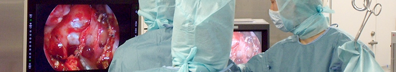
- Treatment of unruptured cerebral aneurysms
Diagnosis of unruptured cerebral aneurysms
Angiography is carried out using MRA, CT angiography, and catheterization
MRA (a test that uses an MRI machine to look at blood vessels) and CT angiography (a test that involves looking at X-ray images taken after receiving an injection of a contrast agent) are carried out for examination. Patients receiving surgical treatment undergo cerebral angiography. A tube called a catheter is inserted via a blood vessel into the groin (the femoral artery) and navigated up as far as the blood vessels in the neck. A chemical called a contrast agent is injected via this catheter, to make the blood vessels stand out on X-rays. This helps identify the relationship between the cerebral aneurysm and the surrounding vessels in detail, and is thus used to choose the best method of treatment and to guide the physician during surgery. As catheterization may cause side effects on rare occasions (such as cerebral infarction as a result of a blood clot), it is only used for patients who require treatment. MRA and CT angiography can be carried out as outpatient procedures, but angiography normally requires a one-night hospital stay for reasons such as the prevention of hemorrhage after catheter removal.
[ Radiological studies for unruptured cerebral aneurysms ]
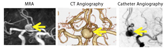
Yellow arrows indicate a cerebral aneurysm.